


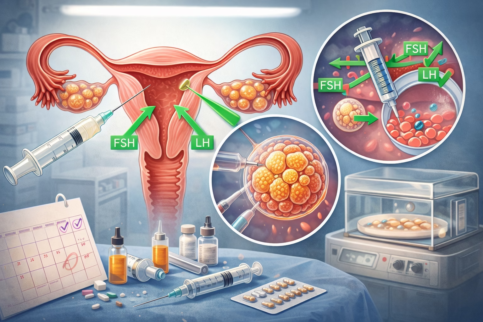


In this article, we will be talking about IVF ultrasound. This technology is an essential part of the in vitro fertilization process. IVF, or in vitro fertilization, is a method used to help those facing fertility challenges to conceive. Ultrasound plays a vital role in this process, allowing healthcare professionals to monitor the development of follicles, assess the lining of the uterus, and guide procedures during treatment. Understanding the significance of IVF ultrasound can help couples navigate their fertility journey more effectively.
IVF ultrasound is a specialized imaging technology that uses sound waves to create visual images of the reproductive organs. It is employed to monitor hormone levels, follicle development, and the overall health of the uterus. During an IVF cycle, monitoring through ultrasound is crucial to ensure the right timing for egg retrieval, embryo transfer, and assessing uterine conditions for implantation. The procedure is non-invasive and helps minimize risks during the treatment process, thereby increasing the chances for a successful pregnancy.
Understanding the Role of IVF Ultrasound
IVF ultrasound essentially assists doctors in keeping track of the stages and progress of the in vitro fertilization process. It provides detailed and real-time images that can aid in decision-making regarding treatment plans. Ultrasound technology is beneficial for monitoring ovarian response to medications, which ultimately determines the timing for egg retrieval. The ultrasound can also help identify any potential complications before they arise.
The ability to visualize internal reproductive organs allows doctors to evaluate factors that can affect fertility treatment. These factors include the size and number of follicles, the thickness of the endometrial lining, and any possible cysts or masses that may need to be addressed. Timing is everything in IVF, and ultrasound plays a key role in ensuring that each step of the process is executed appropriately.
Types of Ultrasound Used in IVF
There are primarily two types of ultrasound used during the IVF process: transvaginal ultrasound and abdominal ultrasound. Each type serves its purpose and offers different advantages.
1. Transvaginal Ultrasound: This method involves inserting a small ultrasound probe into the vagina to obtain clear images of the ovaries and uterus. It is often used during the follicular phase of the IVF cycle when monitoring the development of follicles. The images captured provide information about the size and number of developing eggs, making it easier for the medical team to administer hormone injections effectively.
2. Abdominal Ultrasound: In some cases, abdominal ultrasound may be utilized, especially during the later stages of IVF treatment. It projects sound waves through the abdomen to visualize the uterus and ovaries from a distance. Though less commonly used than transvaginal ultrasound in IVF, it can provide complementary information.
Benefits of Using Ultrasound in IVF
The integration of ultrasound in IVF offers numerous benefits that contribute to better outcomes for patients. Here are some of the key advantages:
- Non-Invasive: Ultrasound techniques are non-invasive, which means they involve minimal discomfort and no surgical intervention.
- Real-Time Monitoring: Doctors can observe the development of follicles in real time, making necessary adjustments to treatment plans when necessary.
- Improved Accuracy: Ultrasound provides accurate measurements of ovarian sizes, enabling precise timing of egg retrieval and embryo transfers.
- Customized Treatment: With detailed imaging, healthcare providers can tailor hormone doses and treatment plans to individual patient needs.
- Reduced Complications: Early detection of issues such as ovarian hyperstimulation syndrome or cyst formation can lead to timely interventions, reducing complications.
How Ultrasound Impacts Egg Retrieval
Egg retrieval is one of the pivotal stages in the IVF process, and ultrasound assistance is key to ensuring that this step is successful. By monitoring follicular development through ultrasound, doctors can closely observe when the follicles reach the ideal size, indicating the readiness of the eggs for retrieval.
A trigger injection of human chorionic gonadotropin (hCG) is typically given when the follicles are mature. After this, the timing for retrieval is critical. Ultrasound enables the medical team to schedule the retrieval procedure at the optimal time, ensuring the maximum number of quality eggs are collected from the ovaries.
The ultrasound is also valuable in guiding the retrieval needle during the procedure. This can significantly minimize risks and help in obtaining eggs without damaging the surrounding tissues.
The Role of Ultrasound in Embryo Transfer
Following egg retrieval, IVF ultrasound is also essential for embryo transfer. Once the eggs have been fertilized and ready to transfer, ultrasound helps position the catheter accurately within the uterus. This ensures that the embryos are placed in the optimal location for implantation, which is crucial for a successful pregnancy.
Having an ultra-clear view of the uterine lining via ultrasound allows the medical team to assess readiness for implantation. A healthy uterine environment is vital for nurturing the embryo. Any abnormalities in the uterine lining can also be evaluated at this time, further informing treatment decisions.
Aftercare and Follow-Up with Ultrasound
After the embryo transfer, monitoring through ultrasound continues to play a crucial role. The initial weeks post-transfer are significant for determining if implantation has occurred. An early pregnancy ultrasound can evaluate the home of the embryo, looking for early signs of pregnancy such as a gestational sac.
Follow-up care is essential in any fertility treatment, and regular ultrasound checks help track the pregnancy progress as it develops. Monitoring can identify any potential complications early on, allowing for timely interventions if necessary. Patients feel more at ease knowing that their doctor is utilizing advanced ultrasound technology to keep track of their health during this delicate time.
Final Thoughts
In summary, IVF ultrasound is an indispensable tool in the in vitro fertilization process. From monitoring ovarian response to guiding egg retrieval and embryo transfers, it enhances patient safety and increases the chances of successful pregnancies. With its non-invasive nature and capacity to provide real-time insights, ultrasound represents an essential facet of modern reproductive medicine.
Patients considering IVF should be aware of the benefits ultrasound offers. Engaging with healthcare professionals who utilize this technology can significantly enhance the overall IVF experience and outcomes. It fosters greater communication between patients and providers and provides clarity amidst the complex landscape of infertility treatments.
For anyone who may undergo this journey, understanding the role of ultrasound helps demystify the process and empowers them in making informed decisions. As technology advances, ultrasound will likely continue to evolve, adding new capabilities that could offer even greater potential in supporting couples on their path to parenthood.
Frequently Asked Questions
Q1: Is IVF ultrasound painful?
A1: IVF ultrasound is generally painless. The transvaginal ultrasound may cause mild discomfort, but it is a short procedure and usually well tolerated.
Q2: How often are ultrasounds performed during IVF?
A2: Ultrasounds may be performed several times throughout an IVF cycle, especially during ovarian stimulation and before egg retrieval.
Q3: Can IVF ultrasound detect problems?
A3: Yes, ultrasound can identify issues such as cysts, fibroids, or abnormal uterine structures, which can impact fertility and treatment planning.
Q4: What if the ultrasound shows no follicles?
A4: The absence of follicles can indicate a need for reevaluation of the stimulation protocols or potential underlying issues that require further investigation.
Q5: How soon after embryo transfer can I have an ultrasound?
A5: Follow-up ultrasounds are typically scheduled about one to two weeks after embryo transfer to check for early signs of pregnancy.
Further Reading
What Type of Psychotherapy Is Best for Anxiety?