


In this article, we will be talking about fetal echo IVF, a crucial procedure that can significantly aid in assessing the health of a fetus at an early stage. Fetal echocardiography, commonly known as fetal echo, is an ultrasound examination specifically designed to evaluate the structure and function of the heart of a fetus. This method is essential for detecting congenital heart defects, which are critical conditions that may require prompt medical attention post-delivery. The integration of IVF, or in vitro fertilization, with fetal echo enhances monitoring and assessing embryo health, making it an important focus for healthcare professionals working with expecting parents.
Fetal echo IVF involves critical monitoring, especially for parents who have a history of heart issues or genetic conditions that could affect a baby’s heart. This examination is typically performed between 18 and 22 weeks of pregnancy, offering detailed images of the baby’s heart chambers, valves, and major blood vessels. The combination of these medical technologies assists doctors in making informed decisions about potential interventions and creating tailored care plans. Understanding the nuances of fetal echo and its role in IVF settings can provide parents peace of mind and better prepare them for the arrival of their newborn.
Understanding Fetal Echo
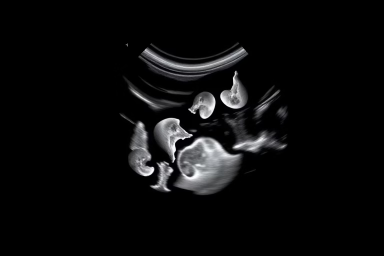
Fetal echo refers to an advanced type of ultrasound that specifically focuses on evaluating the heart of a developing fetus. This detailed assessment is crucial for early detection of congenital heart diseases, which can pose significant health risks. The procedure utilizes sound waves to create real-time images of the fetal heart, allowing healthcare providers to visualize its structure and functionality.
Congenital heart defects occur in approximately 1 in 100 births, making fetal echo an essential tool for expectant parents. The key factors prompting a fetal echo may include family history of heart disease, maternal health concerns, or prior pregnancy complications. During the procedure, a trained technician will apply a gel to the mother’s abdomen and utilize a transducer to capture detailed images. This non-invasive approach yields critical information that can help medical professionals identify potential issues early.
Through this evaluation, specialists can ascertain vital information about the fetal heart, including its size, rhythm, and any abnormalities. This process not only provides insight for immediate medical care but also informs parents of possible outcomes, allowing for better preparation. Fetal echocardiography is now a staple in prenatal care, underscoring its importance in modern obstetrics.
Indications for Fetal Echo IVF
The need for fetal echo IVF typically arises in various circumstances. If parents have a history of congenital heart defects or certain genetic conditions, a fetal echo provides critical insights. A mother’s diabetes or autoimmune disorders can heighten the risk for fetal heart conditions. Other risk factors can include advanced maternal age, prenatal exposure to certain medications, or a family history of other genetic syndromes.
In some cases, abnormal findings from a routine ultrasound may raise concerns requiring further evaluation. These risks underline the need for thorough screening and monitoring throughout pregnancy. By implementing fetal echo in IVF procedures, healthcare providers seek to ensure the safest outcomes for newborns.
- Previous pregnancy with congenital heart disease
- Maternal diabetes or autoimmune disease
- Family history of congenital heart defects
- Cardiac arrhythmias detected in the fetus
- Physical abnormalities identified in routine scans
This list emphasizes the necessity of fetal echo IVF in different contexts, reassuring expectant families that they are receiving comprehensive care for their child’s health.
What to Expect During a Fetal Echo
Undergoing a fetal echo can be a straightforward process, but parents may naturally have apprehensions about the experience. The procedure is typically painless and non-invasive, as it uses ultrasound technology to visualize the fetal heart.
A trained sonographer or cardiologist specializing in fetal echocardiography will perform the examination. The appointment usually takes around 30 to 60 minutes. During the procedure, the technician will place a gel on the mother’s abdomen and then glide a transducer over the skin to capture images of the fetal heart. Parents may be invited to observe the unfolding images, which can be an emotional and hopeful experience.
Once the ultrasound concludes, a specialist will review the findings. In most cases, parents receive reassuring news, but if there are any abnormalities, detailed discussions follow on the potential implications and next steps. Parents may be referred to a pediatric cardiologist or other specialists for further evaluation.
Understanding what to expect during a fetal echo can alleviate anxiety and empower parents to embrace the procedure as a vital step in monitoring their baby’s health.
Benefits of Fetal Echo in IVF
The integration of fetal echo into IVF practices carries considerable benefits. A significant advantage is the early detection of congenital heart defects, which enables healthcare providers to develop effective care strategies. These priorities may include planning for immediate interventions or teaching parents about potential concerns.
The benefits of fetal echo IVF include:
By recognizing potential heart conditions faster, parents and healthcare providers are better equipped to deal with these challenges. Early identification not only contributes to the physical health of the child but also supports families emotionally, allowing them to adjust their expectations and plans as they prepare for their new arrival.
Fetal Echo IVF Procedure: What Happens Next
After receiving results from a fetal echo, the next steps depend primarily on the findings. If the results indicate a healthy fetal heart, parents can move forward confidently through their pregnancy journey. However, abnormal results may lead to a complex healthcare plan involving various specialists.
When heart defects are detected, the healthcare team will provide a comprehensive overview of available options. These may include additional testing, prenatal interventions, or postnatal care plans. Collaborative care often involves consultations with maternal-fetal medicine specialists and pediatric cardiologists, ensuring all aspects of care are addressed.
While the experience of receiving an abnormal diagnosis can be overwhelming for parents, understanding that they are being supported by a dedicated team can offer comfort. Being prepared for postnatal monitoring or interventions can significantly help parents cope as they navigate the complexities following delivery.
Final Thoughts
Fetal echo IVF represents an extraordinary advancement in prenatal care, blending fetal echocardiography and in vitro fertilization to provide comprehensive monitoring for expectant parents. As outlined, the procedure plays a pivotal role in detecting congenital heart defects early, which is critical in crafting timely management plans for impacted infants.
This advanced ultrasounds allow for in-depth evaluations of a fetus’s heart, ensuring families receive the knowledge and preparation necessary for happy, healthy births. The insights gained from a fetal echo can directly affect outcomes, emphasizing the importance of these examinations in contexts where risks are heightened.
Whether through viewing the heart’s activity in real-time or receiving expert evaluations from specialized healthcare providers, the value of fetal echo in the IVF process cannot be overstated. Families are empowered with knowledge while building a comprehensive support system that prepares them for every potential outcome. This makes fetal echo IVF an invaluable part of modern prenatal healthcare.
Frequently Asked Questions
- What is fetal echo IVF and why is it necessary?
Fetal echo IVF involves utilizing echocardiography to assess the health of a fetus’s heart during an IVF pregnancy. It serves as a preventive measure for congenital heart defects.
- When is a fetal echo usually performed?
Typically, fetal echo is conducted between the 18th and 22nd weeks of gestation, though it may be requested sooner based on risk factors.
- Are there any risks associated with fetal echo?
Fetal echo is a non-invasive procedure with minimal risk. It is generally safe for both mother and fetus, as it uses ultrasound technology.
- What should parents expect during the procedure?
Parents can expect a painless experience where a technician applies gel to the abdomen and uses a transducer to visualize and assess fetal heart activity.
- What happens if congenital heart defects are found?
If defects are identified, a detailed plan will be crafted involving specialists, including pediatric cardiologists, to ensure appropriate care before and after delivery.
Further Reading
What Type of Psychotherapy Is Best for Anxiety?