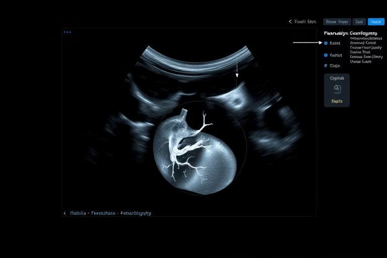
We will be talking about fetal echocardiogram IVF. A fetal echocardiogram is a specialized ultrasound used to assess the heart of a developing fetus. It is an essential tool in prenatal care, allowing healthcare providers to detect potential heart defects early in pregnancy. This procedure is particularly crucial for individuals undergoing in vitro fertilization (IVF), as these pregnancies can sometimes face unique challenges and complications. A fetal echocardiogram provides detailed images of the fetal heart, evaluating its structure, function, and overall health. By identifying any abnormalities early, healthcare providers and parents can prepare and make informed decisions about the pregnancy and potential interventions needed post-birth.
Understanding Fetal Echocardiogram IVF
The process of IVF includes the retrieval of eggs and fertilization outside the body, which can result in a variety of gestational outcomes. The need for a fetal echocardiogram in IVF pregnancies arises from the increased risk of congenital heart defects and other complications. Studies suggest that the likelihood of these conditions may be higher in pregnancies conceived through assisted reproductive technologies. Therefore, acquiring detailed insights into the fetal heart’s condition is vital for ensuring the health of both the mother and the baby.
A fetal echocardiogram evaluates aspects such as heart structure, blood flow, and heart rhythm. This non-invasive technique relies on high-frequency sound waves to produce real-time images of the fetal heart, typically performed around 18 to 22 weeks into the pregnancy. The procedure is quickly becoming a standard part of prenatal care for IVF pregnancies, as timely detection can lead to early management and potentially lifesaving interventions.
Understanding the implications of a fetal echocardiogram can significantly impact the management of a pregnancy, especially for those who have undergone IVF. Knowing that there is an increased risk allows parents and healthcare providers to plan accordingly, ensuring that any detected issues can be addressed effectively. Ultimately, the goal is to provide the best possible outcomes for mothers and their babies through education and proactive healthcare measures.
Why is Fetal Echocardiogram Important?
Fetal echocardiograms are crucial for numerous reasons. They provide vital information about the fetal heart’s structure and function. Detecting heart defects before birth allows parents to prepare for potential treatments immediately after delivery. The following key points highlight their importance:
Healthcare practitioners use echocardiograms to evaluate the anatomy of the heart, including chambers, valves, and vessels. They assess blood flow dynamics to ensure everything functions correctly. In cases of detected abnormalities, obstetricians may refer families to pediatric cardiologists to discuss further evaluation and potential management strategies, such as surgical interventions.
When is a Fetal Echocardiogram Performed?
Typically, a fetal echocardiogram is performed between 18 and 22 weeks of gestation. This specific time frame allows for the fetal heart to be sufficiently developed for detailed imaging. However, in certain circumstances, earlier or later assessments may be recommended:
Following this assessment, further testing or monitoring may be required if concerns arise. Regular follow-ups help ensure the appropriate actions are taken throughout the pregnancy.
Preparation for Fetal Echocardiogram
Preparing for a fetal echocardiogram is straightforward but important. Here are some recommendations for expectant parents to ensure the appointment goes smoothly:
Parents should follow their healthcare provider’s instructions and avoid any measures that might interfere with the imaging results. For example, fasting is generally not required, though hydration is encouraged for better image quality.
It is common to feel excitement or anxiety leading up to the procedure. Remember, this is a routine part of prenatal care designed to ensure the wellbeing of both mother and baby.
What to Expect During the Procedure?
A fetal echocardiogram typically takes 30 to 60 minutes. Expect to be in a quiet, comfortable room for the procedure. A trained technician or healthcare provider will conduct the scan, following these steps:
Throughout the process, it is essential to remain still and follow any instructions given by the technician. Parents may also hear the fetal heartbeat during the exam, providing reassurance.
After the Fetal Echocardiogram
After a fetal echocardiogram, the relevant healthcare provider will share the findings of the assessment. Here are a few common experiences:
Recovery from the procedure is immediate, as it is non-invasive, allowing parents to resume normal activities right away. Engaging in discussions regarding the results with your healthcare provider can provide peace of mind and ensure that you are prepared for any necessary actions moving forward.
Final Thoughts
In summary, the fetal echocardiogram plays a significant role in monitoring pregnancies, specifically those conceived through IVF. Through early detection of potential heart defects, this procedure allows for timely decision-making, intervention, and management. Understanding why these tests are necessary, when to have them, and what to expect can help ease concerns for expectant parents and prepare them for various outcomes. A proactive approach is essential in these pregnancies, as it increases the chance of favorable results.
Parents should engage in open discussions with healthcare providers about their specific circumstances, including any family history of congenital heart defects, maternal health issues, or notable findings in standard ultrasounds. Ultimately, the goal remains to prioritize the health and wellbeing of both mother and child.
Frequently Asked Questions
1. What is the primary purpose of a fetal echocardiogram?
The primary purpose is to assess the fetal heart’s structure and function, identifying any potential congenital heart defects early in the pregnancy.
2. How is a fetal echocardiogram different from a regular ultrasound?
A fetal echocardiogram specifically focuses on the heart, utilizing advanced techniques to evaluate the heart’s anatomy and function, unlike routine ultrasounds that provide overall fetal imaging.
3. Is a fetal echocardiogram safe?
Yes, it is a safe and non-invasive procedure that uses sound waves to create images and poses no risk to the mother or fetus.
4. Can I find out the sex of my baby during a fetal echocardiogram?
While the primary focus is on the heart, the technician may occasionally identify the baby’s sex if viewed during the procedure.
5. Will I need any special preparations for the fetal echocardiogram?
No special preparations are needed, but it is advisable to stay hydrated, and comfortable clothing is recommended for ease of access.
Further Reading
What Type of Psychotherapy Is Best for Anxiety?


