
Today we will be talking about infections and their visibility on a PET scan. A PET (positron emission tomography) scan is a medical imaging technique that allows healthcare professionals to observe metabolic processes in the body. It is commonly used to detect cancer and assess heart and brain conditions. Infections can also appear on PET scans, but understanding how they do requires a closer look at both the nature of infections and the technology behind PET imaging.
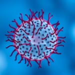
An infection is when harmful microorganisms, such as bacteria, viruses, fungi, or parasites invade the body, multiply, and begin to damage tissues and disrupt normal biological functions. Infections can vary greatly in their severity and the symptoms they produce, ranging from mild disturbances to life-threatening illnesses. The immune system typically responds to infections with inflammation, which can cause various clinical manifestations like fever, pain, and swelling. In modern medicine, infectious diseases are diagnosed through clinical evaluation, laboratory tests, and imaging techniques like PET scans. When it comes to PET scans, the detection of an infection often relies on the application of a radioactive tracer, mainly fluorodeoxyglucose (FDG), which reflects glucose metabolism associated with heightened biological activity, such as inflammation or infection.
1. How PET Scans Work
The process behind PET scans utilizes a radioactive tracer that emits positrons as it decays. The most common tracer is fluorodeoxyglucose (FDG), which is similar to glucose and allows for mapping areas of the body that have higher metabolic activity. When an infection occurs, the body’s immune response increases blood flow and glucose uptake in the affected tissues. When the PET scan captures this response, the infection can be seen as an area of increased uptake on the imaging results.
This modality effectively highlights regions of abnormal activity, making it a valuable tool for diagnosing not just cancer but infections as well. The detailed information provided by PET scans can help clinicians determine the location, extent, and even type of infection. Moreover, this technique can differentiate between infection and other conditions such as tumors or inflammation due to other causes, enhancing the diagnostic process.
2. Types of Infections Visible on PET Scans
Infections can be broadly categorized into bacterial, viral, fungal, and parasitic infections, and many of these can be visualized on a PET scan. Bacterial infections, for instance, can arise in different body parts, including bones (osteomyelitis), lungs (pneumonia), or soft tissue. *The intensifying metabolic activity associated with bacterial growth and the subsequent immune response often render these infections visible on the scan.*
Viral infections may also appear on PET scans; however, visualization depends heavily on the body’s inflammatory response. Fungal infections, such as those caused by Aspergillus or Cryptococcus, can show up due to the body’s immune activity. Furthermore, parasitic infections like those caused by Toxoplasma Gondii can also be identified on PET scans, particularly when they provoke inflammatory responses. Understanding the type of infection is crucial for selecting appropriate treatment strategies and monitoring the condition’s progression.
3. Importance of Early Detection
Catching an infection early can significantly impact treatment outcomes and limit complications. Utilizing PET scans allows for prompt identification and localization of infections that might not be apparent in other imaging modalities such as X-rays or MRIs. Early detection can facilitate quicker response times for antibiotics, antivirals, or other necessary interventions. Not only does this improve outcomes for patients, but it also contributes to more efficient healthcare management.
In the case of severe infections or abscess formations, the ability to visualize these conditions through a non-invasive procedure like a PET scan minimizes the need for exploratory surgery. This aspect is particularly advantageous for patients who may be at higher surgical risk or those with multiple health conditions. Therefore, integrating PET scans in clinical settings becomes an immensely valuable asset in infectious disease management.
4. Comparing PET Scans to Other Imaging Techniques
When considering the diagnosis of infections, other imaging methods, such as CT (computed tomography) and MRI (magnetic resonance imaging), can also play a role. Each modality has its strengths and weaknesses, leading to specific applications based on clinical scenarios.
CT scans are often used for evaluating infections in the lungs, sinus cavities, and abdominal organs, offering excellent detail on anatomical structures. On the other hand, MRI is particularly useful for assessing infections of the central nervous system or soft tissues, as it provides superior contrast resolution between different types of soft tissues.
However, PET scans have distinct advantages in assessing metabolic activity. While the other modalities provide structural imaging, PET scans directly visualize the body’s metabolic processes, enabling better distinction between benign and malignant conditions as well as active infections. Additionally, PET/CT machines combine both imaging types for integrated insights, ultimately enhancing the precision of diagnosis.
5. Challenges in Identifying Infections on PET Scans
While PET scans are beneficial in identifying infections, certain challenges may arise. Not all infections will appear distinctly, especially if the body’s immune response is minimal. For example, a chronic infection may not produce significant metabolic changes while still causing damage. Similarly, certain cancers may mimic infections in PET imaging, posing diagnostic dilemmas. High levels of inflammation from conditions like vasculitis or autoimmune diseases can also lead to false positives.
Striking a balance between leveraging the benefits of PET scanning and being aware of its limitations is paramount. Clinicians must interpret PET scan results in conjunction with clinical presentation and corroborative diagnostic tools. This holistic approach enhances accurate diagnosis and appropriate treatment plans.
6. The Role of Radiologists in Diagnosing Infections
Radiologists play a crucial role in the interpretation of PET scans and other imaging techniques. Their expertise is essential for distinguishing active infections from other potential conditions that may appear on imaging. Radiologists analyze the size, shape, location, and metabolic activity of areas in question, synthesizing this data with the patient’s clinical history.
Their analysis provides valuable insights for treating physicians, aiding them in making informed decisions about follow-up tests, interventions, or specific treatments. Effective communication between radiologists and treating doctors enhances patient care and ensures timely management of infections, ultimately improving outcomes and minimizing complications.
7. The Future of PET Scanning in Infectious Disease
As technology continues to advance, PET scanning is becoming increasingly sophisticated, allowing for enhanced imaging capabilities and better diagnostic accuracy. Upcoming innovations may include targeted tracers that differentiate between types of infections or even more advanced imaging algorithms that decrease false positives and negatives.
Furthermore, combining PET scanning with other emerging technologies, such as artificial intelligence, may refine diagnostic capabilities. AI can analyze large datasets to identify patterns associated with infections, improving accuracy in interpretation and perhaps even providing early warning systems for outbreaks.
8. Treatment Implications Based on PET Scan Results
The results from PET scans can significantly influence treatment choices. For instance, understanding the extent of an infection can affect decisions on the necessary duration of antibiotic treatment, surgical interventions, or even the need for additional diagnostics.
Additionally, if a scan reveals an infection resistant to typical treatments, healthcare providers may pivot to more aggressive therapy or alternative medications. Monitoring treatment effectiveness through follow-up PET scans can also reveal how well a patient is responding to therapy. This approach ensures that healthcare providers can adapt strategies in real-time based on successful or unsuccessful treatment responses.
9. Patient Experience and PET Scans
For patients, undergoing a PET scan can evoke feelings of anxiety or uncertainty about what the process entails. Education about the procedure can help alleviate concerns. Typically, the process lasts about two hours, during which the patient will receive the radioactive tracer, wait for absorption, and then undergo the scan itself.
Comfort measures, such as relaxing environments and explanations from healthcare professionals, can enhance the patient experience. Understanding the significance of the scan can empower patients and lead to increased cooperation during the imaging process. Furthermore, clear communication about potential results and what they mean fosters trust between patients and healthcare providers.
10. Common Misconceptions about PET Scans
Despite the utility of PET scans, numerous misconceptions persist, which can lead to confusion or anxiety. Many people mistakenly believe that PET scans can definitively diagnose all types of infections or conditions. In reality, while PET scans provide critical information, they must be interpreted alongside clinical evaluations and other diagnostic tests.
Another common misconception is that PET scans are highly harmful due to radiation exposure. The amount of radiation administered is relatively low and is comparable to that of other diagnostic imaging procedures. Educating patients and the public about the benefits and limitations of PET scans can promote a better understanding of their role in diagnosing infections and other medical conditions.
In conclusion, PET scans are a vital tool in diagnosing infections, demonstrating that heightened metabolic activity associated with infections can be visualized effectively. Understanding how PET scans work, the types of infections visible on these scans, the importance of early detection, and the role radiologists play enhances the management of infectious diseases. With advancements in technology, the capabilities of PET scans continue to evolve, promising even more accurate diagnostic pathways in the future. By addressing challenges and misconceptions surrounding PET scans, healthcare providers can better serve their patients and ensure that they receive precise diagnoses and appropriate treatments.
Frequently Asked Questions
1. Can all infections be seen on a PET scan? Not all infections are visible on a PET scan. While many bacterial and fungal infections can be identified due to increased metabolic activity, some chronic conditions may not produce significant changes.
2. Are PET scans safe? Yes, PET scans are generally safe and involve minimal radiation exposure, comparable to other imaging techniques. The benefits of the scan often far outweigh potential risks.
3. How long does a PET scan take? A PET scan usually takes about two hours, including time for the radioactive tracer to be absorbed and the scan itself.
4. Will a PET scan hurt? A PET scan is non-invasive and typically does not cause pain. Patients may feel a small pinch during the injection of the tracer.
5. What should I do to prepare for a PET scan? Preparation varies by individual. Typically, patients are advised to fast for several hours before the scan to avoid any interference with the results. Following specific instructions provided by healthcare professionals is essential.
Further Reading
3.5 tog sleeping bag temperature guide
What Type of Psychotherapy Is Best for Anxiety?