Key Takeaways
- The maxillary sinuses are located directly above the upper back teeth.
- Root displacement can occur during dental extractions or due to bone loss.
- Common symptoms include sinus pressure, nasal congestion, and facial pain.
- Professional imaging and surgical intervention are often required for removal.
Understanding the Relationship Between Your Teeth and Sinuses
When you experience facial pain or discomfort, it is not always immediately clear whether the problem originates from your teeth or your sinuses. A specific concern involves tooth root in maxillary sinus symptoms, which refers to the displacement of a tooth root or a fragment into the sinus cavity located just above your upper molars. Because the roots of these teeth are often separated from the sinus by only a very thin layer of bone, any structural issue can lead to complex symptoms that require careful evaluation.
The maxillary sinuses are air-filled spaces positioned behind your cheekbones. When a tooth root enters this space, it can trigger localized irritation, inflammation, or infection. Understanding how this happens and how to recognize the signs is an essential part of maintaining your oral and respiratory health. While this condition sounds intimidating, modern dentistry and oral surgery offer effective ways to diagnose and resolve the issue safely.
This guide explores the underlying causes of root displacement, the specific indicators you should look for, and the diagnostic processes used by professionals. Please remember that this information is for educational purposes only. If you suspect you have a problem, you must consult with your dentist or an oral surgeon to receive a proper diagnosis and treatment plan tailored to your specific situation.
Anatomy and Causes of Root Displacement
To understand why tooth root in maxillary sinus symptoms occur, we must first look at the anatomy of the upper jaw. The maxillary sinuses, also known as the antrum of Highmore, are the largest of the paranasal sinuses. The roots of the upper premolars and molars are often in close proximity to the floor of these sinuses. In some individuals, the bone separating the tooth root from the sinus is extremely thin or even absent, leaving only a membrane between the two.
Root displacement typically occurs during routine dental procedures, most commonly during the extraction of an upper molar. If the bone surrounding the root is fragile or if the root is particularly long and curved, it may accidentally be pushed into the sinus cavity rather than being removed through the socket. This is a known complication that dentists are trained to anticipate and manage.
Beyond extractions, other factors can contribute to this issue. Chronic periodontal disease can erode the bone that normally acts as a barrier, making the area more vulnerable. Additionally, traumatic injuries to the face can cause fractures in the alveolar bone, potentially shifting tooth fragments into the sinus. Identifying these risks early helps dental professionals take extra precautions during surgeries to prevent these complications from occurring.
Recognizing Tooth Root in Maxillary Sinus Symptoms
The symptoms associated with a displaced tooth root can often mimic common sinus infections or dental abscesses, making them tricky to identify without professional imaging. Patients may report a persistent feeling of pressure or fullness in the cheek area that does not respond to standard decongestants or antibiotics. This discomfort is often localized to one side of the face, corresponding to the tooth that was extracted or injured.
One of the most telling signs is a change in nasal symptoms. You might notice a foul-smelling or bad-tasting discharge from one nostril. This occurs because the presence of a foreign object, such as a root fragment, can block the normal drainage of the sinus, leading to fluid buildup and potential bacterial growth. If you notice persistent congestion that refuses to clear up weeks after a dental procedure, it is a significant red flag.
Other common indicators include:
- Persistent facial pain that increases when bending forward or lying down.
- A sensation of air or fluid escaping through the tooth socket when you blow your nose.
- Increased sensitivity or pain in the upper teeth near the site of the extraction.
- Visible swelling or tenderness in the gum tissue surrounding the area of the previous tooth.

Diagnostic Procedures and Imaging
When you present with potential tooth root in maxillary sinus symptoms, your dentist will need to visualize the area clearly. A standard physical examination is rarely sufficient because the root fragment is hidden deep within the sinus. Instead, your doctor will rely on advanced imaging technology to pinpoint the exact location, size, and orientation of the displaced fragment.
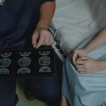
The most common diagnostic tool is a periapical X-ray, which provides a detailed look at the tooth socket and immediate surrounding bone. However, for a more comprehensive view, a Cone Beam Computed Tomography (CBCT) scan is often preferred. A CBCT scan provides a three-dimensional image of your jaw, teeth, and sinuses, allowing the surgeon to see exactly where the root fragment is resting and whether it has caused any secondary issues like sinus membrane inflammation.
| Imaging Method | Primary Benefit |
|---|---|
| Periapical X-ray | Quick, low radiation, good for initial screening |
| Panoramic X-ray | Provides a broad view of the entire jaw and sinus area |
| CBCT Scan | High-resolution 3D imaging for precise surgical planning |
Treatment Options for Displaced Roots
Once a diagnosis is confirmed, the primary goal is the safe removal of the foreign object. Leaving a tooth root in the sinus can lead to chronic sinusitis, which is an ongoing inflammation of the sinus lining. Treatment usually involves a procedure called an antrostomy or a functional endoscopic sinus surgery (FESS), depending on the location of the root fragment and the status of your sinus health.
In some cases, if the root is near the extraction site, the surgeon may be able to retrieve it through the original tooth socket. This is often the least invasive approach. If the fragment has moved deeper into the sinus, a small incision may be made in the gum above the teeth to access the sinus floor, allowing for the direct removal of the object. This is typically performed by an oral and maxillofacial surgeon under appropriate anesthesia.
Following the removal of the fragment, the surgeon will ensure that the site is cleaned thoroughly. If an opening between the sinus and the mouth was created, known as an oro-antral communication, the surgeon will take steps to close it. This might involve using a small flap of gum tissue or other specialized materials to ensure the area heals properly and prevents future sinus issues.

Recovery and Post-Operative Care
Recovery after the removal of a tooth root from the sinus requires patience and adherence to your surgeon’s instructions. You will likely be prescribed a period of rest, and you may be advised to avoid any activities that increase pressure in your sinus cavities. This includes blowing your nose forcefully, sneezing with your mouth closed, or using straws, as these actions can disrupt the healing tissue.
Pain management is a standard part of post-operative care. Your doctor will provide guidance on how to manage any discomfort using over-the-counter or prescription medications. Keeping your head elevated while sleeping can also help reduce swelling and facial pressure. It is common to experience some congestion for a few days, but this should gradually subside as the sinus lining heals.
Maintaining good oral hygiene is equally important, though you must be careful around the surgical site. Your dentist may recommend a specific mouth rinse to keep the area clean without causing irritation. Regular follow-up appointments are essential to ensure the sinus is healing correctly and that there are no signs of lingering infection. Most patients recover fully within a few weeks, provided they follow the prescribed aftercare routine.
Frequently Asked Questions
Can a tooth root in the sinus cause a headache?
Yes, it can. Because the maxillary sinuses are located near the nerves that supply the face, inflammation or pressure caused by a foreign object can manifest as a persistent, dull headache or facial pain that feels like a tension headache.
Is it always necessary to remove a tooth root from the sinus?
Generally, yes. A tooth root is considered a foreign body. If left in the sinus, it can act as a nidus for bacteria, leading to chronic infections, sinus inflammation, and other long-term complications that are much harder to treat than the initial removal.
How long does the surgery take?
The duration of the surgery depends on the complexity of the case, such as the size and location of the fragment. Most procedures are relatively quick, often taking less than an hour, but the total time may vary depending on the anesthesia used and the specific surgical approach required.
Will I need antibiotics after the procedure?
It is common for surgeons to prescribe a course of antibiotics to prevent or treat potential sinus infections, especially if there was already evidence of inflammation or if the procedure involved entering the sinus cavity. Always follow the full course as directed by your healthcare provider.
The Bottom Line
Dealing with tooth root in maxillary sinus symptoms can be an unsettling experience, but it is a manageable condition with the right professional care. By recognizing the signs early, such as persistent unilateral facial pressure or unusual nasal discharge, you can seek the help you need before the issue progresses to a chronic infection. Modern imaging techniques like CBCT scans make diagnosis accurate, while oral surgeons possess the expertise to remove the fragment safely and effectively. If you are experiencing these symptoms, prioritize an appointment with your dentist to ensure your long-term health and comfort.